**** Human Pinealocytes ****
For more detail, please refer to the published papers.
1. Hasegawa, A., Ohtsubo, K., Izumiyama, N., and Shimada, H.: Ultrastructural study of the human pineal gland in aged patients including a centenarian. Acta Pathol Jpn, 40; 30-40, 1990 (pdf 1.3MB available).
2. Hasegawa, A., Shimada, H., Izumiyama, N., and Ohtsubo, K.: Paired twisted filaments in human pinealocytes. Acta Pathol Jpn, 41; 265-269,1991 (pdf 5.0MB available).
1. Light microscopic features of the human pinealocytes
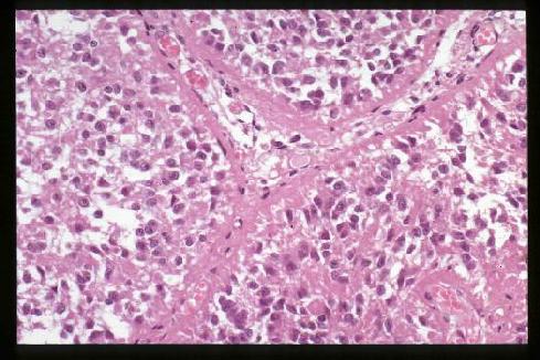
Human pinealocytes
Human pinealocytes in alveolar arrangement (H&E stain). The appearance
of the pineal gland is fairly different even within the mammalian,
not to mention the dramatic morphological changes in the process of the evolution of
vertebrates from fish to human. The phylogenesis of the pineal may be more
interesting than Much Melatonin About Nothing.
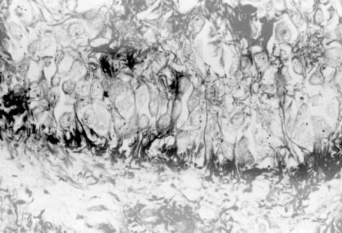
Rio-Hortega stain
Bulbous endings of human pinealocytes in contact with
the intervening capillary stroma (lower half)(Rio-Hortega stain)
2. Electron microscopic features of the human
pinealocytes
AUTHOR'S NOTE: Although the specimens were taken at autopsy within 5 hours of death
(mean 2.5 hours), the post mortem degeneration of membranous ultrastructures
cannot be ignored in the electron micrographs of human materials.
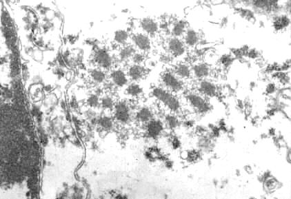
Synaptic ribbons in aged human
Electron micrograph of an aged human pineal gland, showing a synaptic
ribbon field composed of many synaptic ribbons in the close vicinity of
the plasma membrane. 100-year-old female (original x25,000). On the
physiological significance of the synaptic ribbon, refer to the corresponding
page (Pineal gland of rodent species) and other
references (e.g., Vollrath L: Synaptic ribbons of a mammalian pineal gland;
Circadian changes. Z Zellforsch, 145; 171-183, 1973)
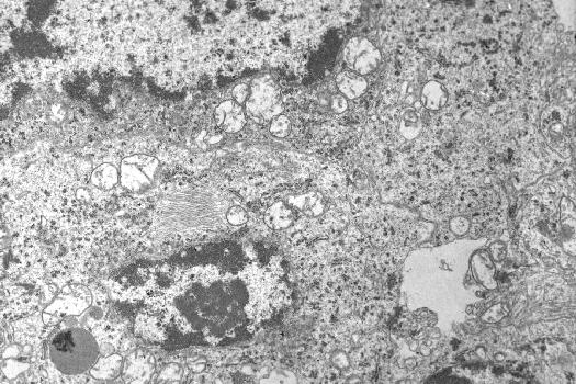
Paired twisted filaments in a human pineal parenchymal cell
(in the center left, original x9,300)
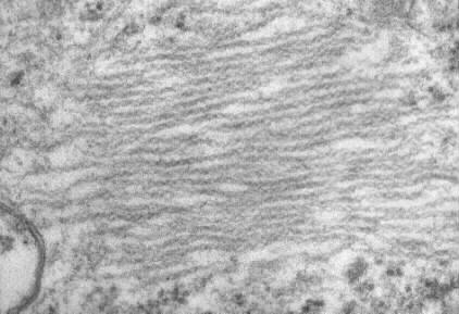
Enlarged view
Enlarged longitudinal section of the paired twisted filaments
(PTF), peculiar double helical structures, 12 to 25 nm in maximal helix
width with a half periodicity of 30 to 35 nm (periodicity of the constrictions),
in a human pinealocyte (original x74,000). These PTF were first
reported in human pinealomas by Hassoun et al (Acta Neuropathol, 65;
163-165, 1984) and similar structures have been described only exceptionally
in spinal ganglia (van den Bosch et al, in Virchows Arch [Cell Pathol],
47; 217-222, 1984) and cerebral neurons (Knox et al, in Acta Neuropathol,
52; 7-15, 1980) of aged rats. The biological and pathological implications
of the PTF remain unclear.
Back to previous Home Page at AOL (Till October 31 2008)
Back to previous Home Page at geocities.jp web host (Till March 31 2019)
Back to New Home Page (Translocated Sakura server)
-----------
Copyright 1997-2010, Akio Hasegawa & the Japanese Society of Pathology
version 1.30 2010/6/26
-----------
Please send your comments to DrHASEGAWA@aol.com